
appendicular skeleton Diagram Quizlet
Appendicular Skeleton (126 bones) | SEER Training. Home » Cancer Registration & Surveillance Modules » Anatomy & Physiology » Skeletal System » Divisions of the Skeleton Appendicular Skeleton (126 bones)

Appendicular Skeleton Chart The Chart
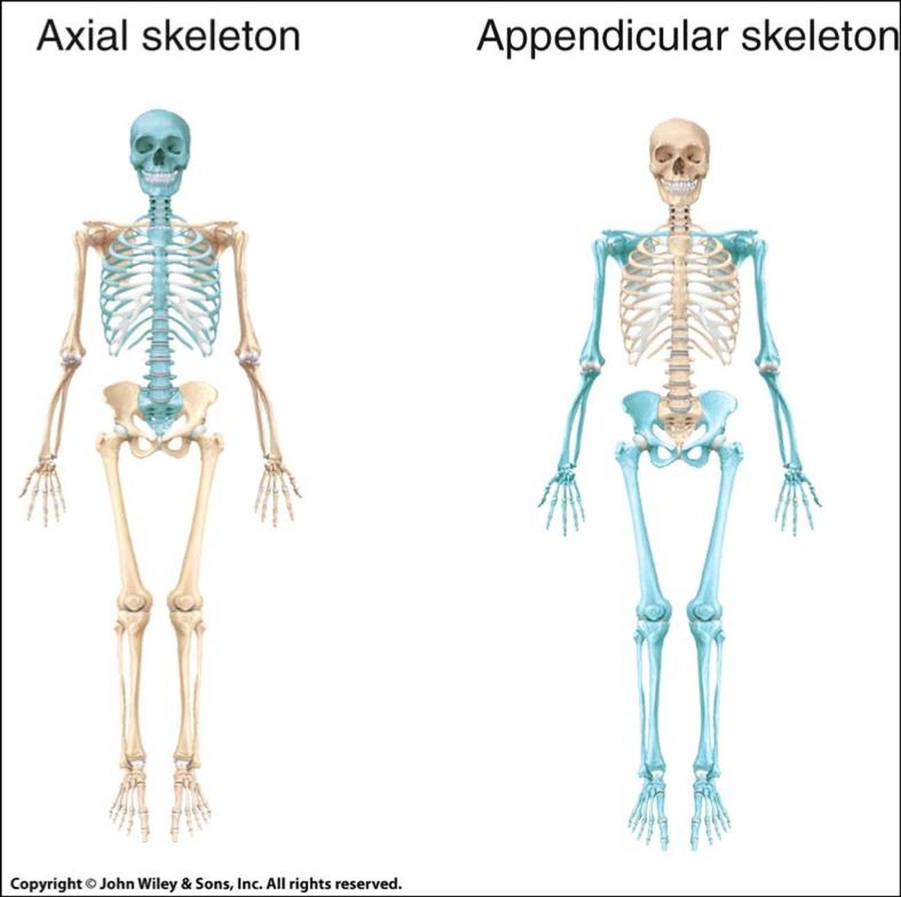
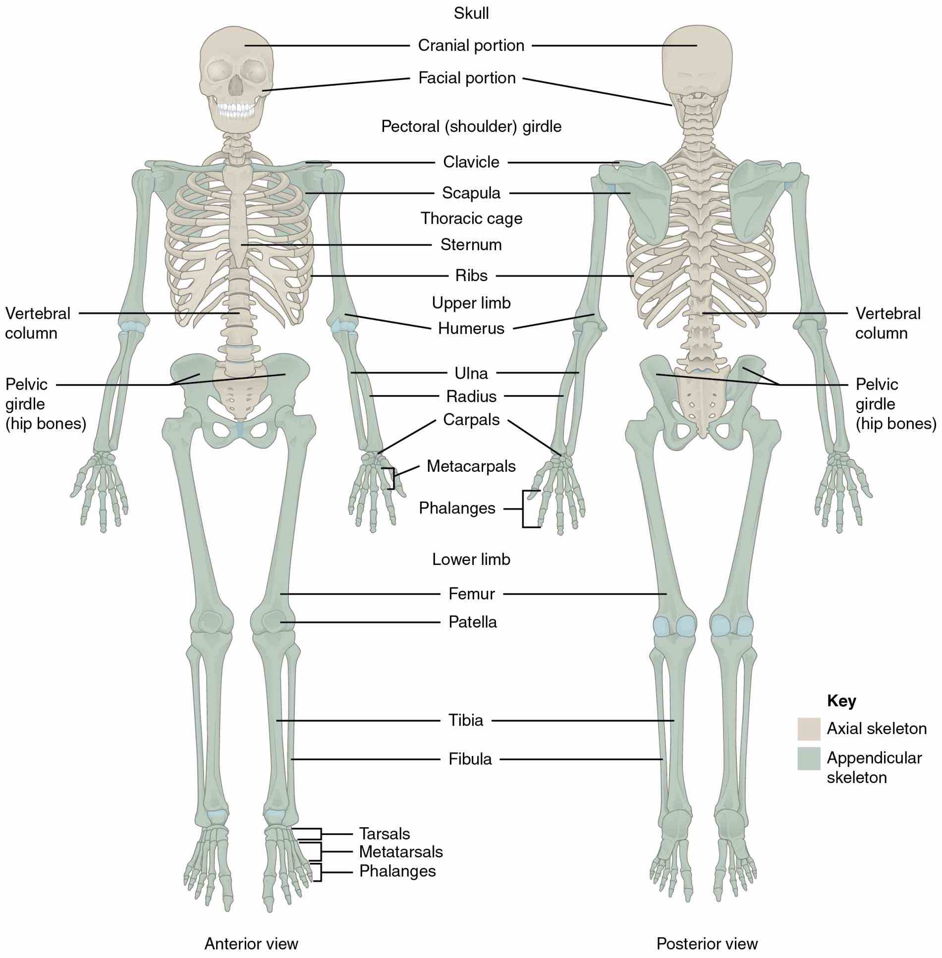
The axial skeleton comprises the bones found along the central axis traveling down the center of the body. The appendicular skeleton comprises the bones appended to the central axis. Above: The bones of the axial skeleton make up the central axis of the body including the skull, hyoid, vertebrae, ribs, sternum, sacrum, and coccyx.

Axial and Appendicular Skeleton
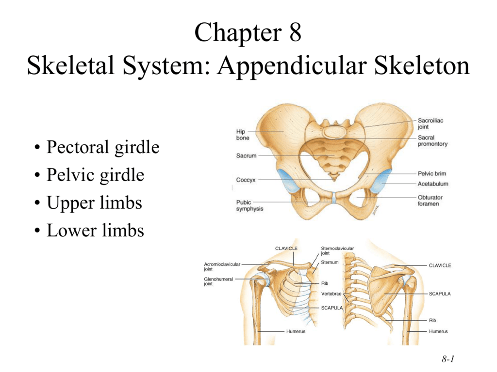
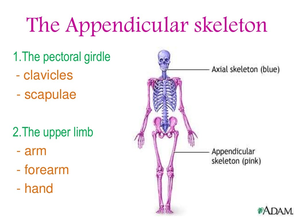
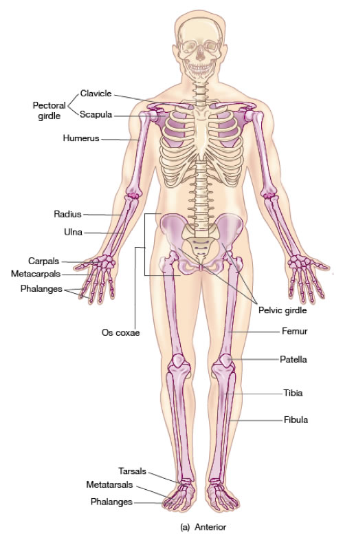
1. The Bones of the Shoulder Girdle The pectoral or shoulder girdle consists of the scapulae and clavicles. The shoulder girdle connects the bones of the upper limbs to the axial skeleton. These bones also provide attachment for muscles that move the shoulders and upper limbs. See the bones of the shoulder girdle in 3D: 2. Bones of the Upper Limbs

Appendicular Skeleton These bones are part of the human
Key Terms. Girdle: A group of bones that connect the appendages to the axial skeleton.; phalanges: The digital bones of the hands and feet (singular, phalanx).; appendages: The parts of the body that extend from the axial trunk.; The appendicular skeleton of vertebrates, including humans, consists of the bones that support and compose the appendages (for example, the arms and legs of humans).
INTRODUCTION TO THE APPENDICULAR SKELETON
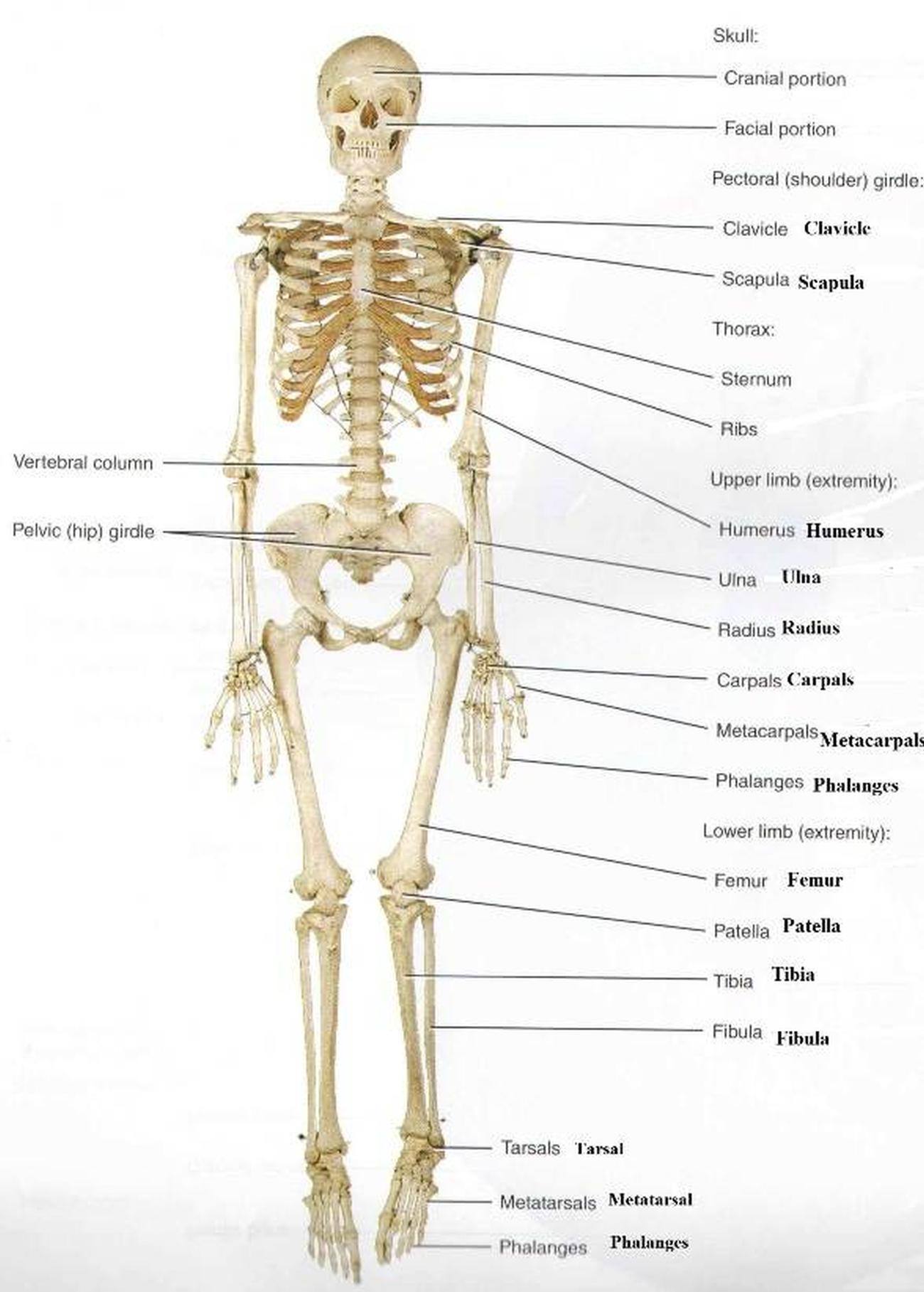
Part 1: Review of the Appendicular Skeleton. Pectoral/Shoulder Girdle. Upper Limb. Pelvic Girdle. Lower Limb. Part 2: Laboratory Activities - Pectoral/Shoulder Girdle & Upper Limb. Pectoral/Shoulder Girdle. Clavicle - Anatomical left from anatomical right. Scapula - Fill in four steps to determine anatomical left from anatomical right.

Image result for axial skeleton anatomy labeled Anatomy bones
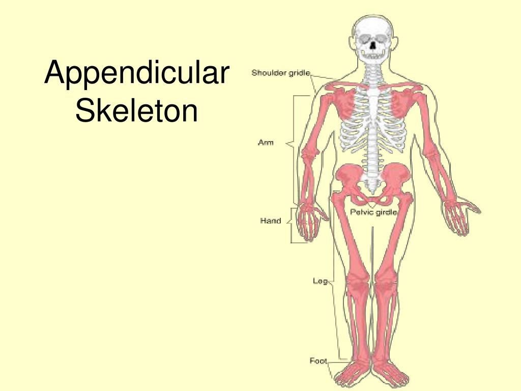
The appendicular skeleton is made up by the bones attached or appended to the axial skeleton. These are the bones of the limbs, hands, and feet, the bones of the pectoral (shoulder) girdles, and the coxal bones of the pelvic girdle. Figure \(\PageIndex{1}\): The appendicular skeleton highlighted in blue.
Pictures Of Appendicular Skeleton
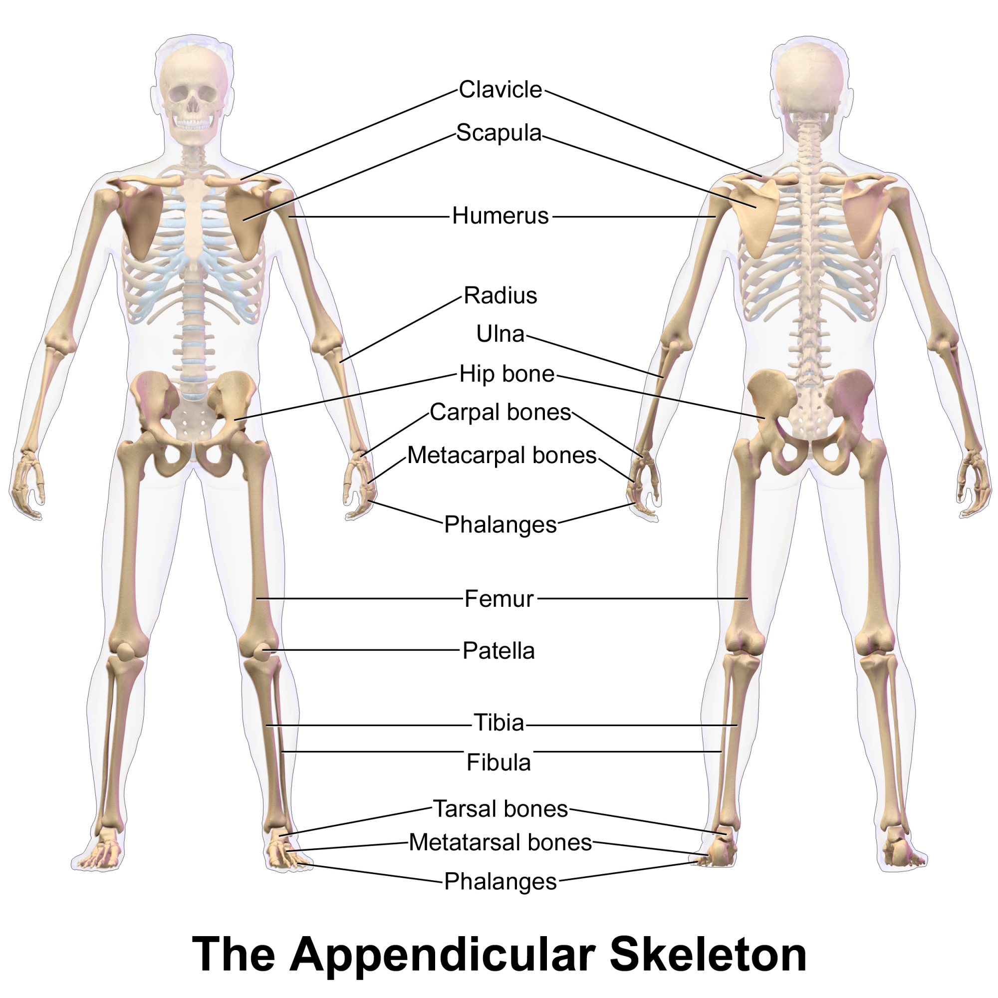
The appendicular skeleton is the portion of the skeleton of vertebrates consisting of the bones that support the appendages. There are 126 bones. The appendicular skeleton includes the skeletal elements within the limbs, as well as supporting shoulder girdle and pelvic girdle. [1]
The appendicular skeleton
Muscles that position the pectoral girdle are located either on the anterior thorax or on the posterior thorax (Figure 10.6.1 10.6. 1 and Table 10.6.1 10.6. 1 ). The anterior muscles include the subclavius, pectoralis minor, and serratus anterior. The posterior muscles include the trapezius, rhomboid major, and rhomboid minor.

Appendicular Skeleton Learn Skeleton Anatomy
6 - the appendicular skeleton: learn the bones the arms and legs. 7 - the heart: name the parts of the bones. 8 - the foot: Can you identify the bones of the foot? 9 - anatomy terminology: Do you know the language of anatomy? 10 - the skeleton, posterior: identify the skeleton from behind. Anatomy Physiology Therapies Resources Faq
The appendicular skeleton of human body Online Science Notes
The appendicular skeleton is one of the two major groups of bones in the human skeleton. It consists of the bones of the limbs (or appendages), and the bones that attach the limbs to the rest of the body. It includes a total of 126 bones, including those in the arms, legs, and shoulder and pelvic girdle bones.
PPT Appendicular Skeleton PowerPoint Presentation, free download ID
The appendicular skeleton is one of two major bone groups in the body, the other being the axial skeleton. The appendicular skeleton is comprised of the upper and lower extremities, which include the shoulder girdle and pelvis.

Skeletal System Introduction , Parts, Functions, Diagram & Fact
Labeling Exercises: Crossword Puzzles: Flashcards: Concentration: Internet Activities: Chapter Weblinks. Help Center: Human Anatomy, 6/e. Kent Van De Graaff, Weber State University. Skeletal System: The Appendicular Skeleton. Labeling Exercises. Skeleton-Anterior View Skeleton-Posterior View Lower Skeleton Upper Skeleton-Anterior View Upper.
Anatomy Of Appendicular Skeleton Bones
Labeled pictures of appendicular skeleton Students also viewed Appendicular Skeleton Teacher 134 terms Reese_Beltman Preview The Appendicular Skeleton 105 terms Kaela_Valdez Preview Bones and Bone Markings: The Appendicular Skeleton 92 terms gavin_spennewyn Preview Appendicular Skeleton Teacher 41 terms Mrs_M_Stewart Preview Appendicular Skeleton

PPT The Skeleton is divided into 2 parts PowerPoint Presentation
Appendicular Skeleton Labeling by mcdonald.lara2 143,346 plays 16 questions ~40 sec English 16p More 3.93 (you: not rated) Tries Unlimited [?] Last Played November 26, 2023 - 08:23 PM There is a printable worksheet available for download here so you can take the quiz with pen and paper. Remaining 0 Correct 0 Wrong 0 Press play! 0% 0:00.0
Skeletal System Appendicular Overview
Label the Bones of the Appendicular Skeleton As part of the skeletal system unit, anatomy students explore each bone and their structures. For example, each group is given a femur (I have a collection of real and plastic models) and label structures. Structures include the trochanters, condyles, head, neck, and linea aspera.
Pictures Of Appendicular Skeleton
The appendicular skeleton contains the bones of the upper and lower limbs (arms and legs). What is the Appendicular Skeleton? The human body is comprised of roughly 206 to 213 different bones..