
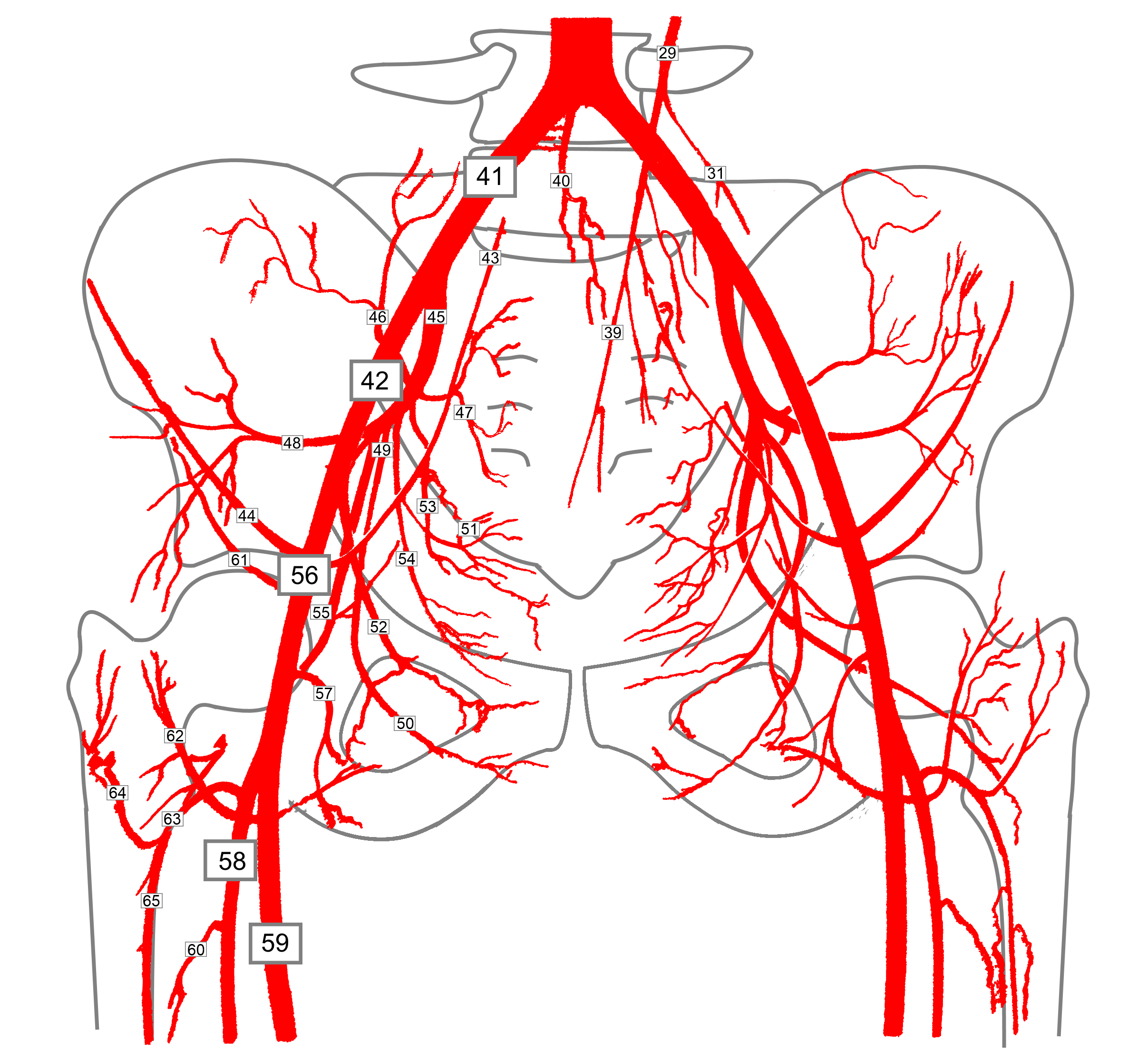
Iliac Artery Common iliac artery, Internal & External iliac artery branches
Isolierte Iliakaaneurysmen sind eine seltene Form aortoabdominaler Aneurysmen und weisen eine hohe Komplikations- und Rupturrate auf, die mit einer hohen Mortalität einhergeht. Unspezifische Symptome verzögern die Diagnosestellung.

How to Pronounce Vena iliaca? YouTube
a iliaca communis dextra et sinistra. unpaired continuation of aorta. a sacralis mediana. branches of aorta ascendens. a coronaria dextra, a coronaria sinistra. bulbus aortae is located at: 3rd left rib (origin) bulbus aortae is made up of. 3 sinus aortae.
Surgery Assistant
The common iliac artery is a large artery of the abdomen paired on each side. It originates from the aortic bifurcation at the level of the 4th lumbar vertebra. It ends in front of the sacroiliac joint, one on either side, and each bifurcates into the external and internal iliac arteries . Structure
Arteria Iliaca communis Anatomie, Verlauf, Äste & pAVK Kenhub
Verlauf. Die Arteria iliaca communis ist beim Erwachsenen nur etwa 3-4 cm lang, hat aber einen Durchmesser von mehr als 10 mm. Nach kurzem Verlauf teilt sie sich in der Nähe des Iliosakralgelenks in zwei Äste auf: Die Arteria iliaca communis wird von der gleichnamigen Vena iliaca communis begleitet.
Arteria Iliaca communis Anatomie, Verlauf, Äste & pAVK Kenhub
Iliac arterial aneurysms are focal dilatations of the iliac artery . Although the dimensions that define the aneurysm are dependent on the sex of the patient and the portion of the artery involved, a common iliac artery (CIA) with a diameter ≥1.7 cm in males or ≥1.5 cm in females is considered ectatic. A diameter >2.5 cm is considered aneurysmal 3.

AMIGOS PARA SIEMPRE Anatomía humana Arterias
10.01.2022 Udvidelse af bækkenpulsåren (a. iliaca aneurisme) Røntgenkontrastundersøgelse (arteriografi) af legems- og bækkenpulsårerne (aorta og iliaca arterierne). I højre bækkenpulsåre (arteria iliaca communis) ses en stor udposning (aneurisme) 1 Aneurisme (udposning) Kateter højre a. iliaca interna/hypogastrica højre a. iliaca externa Aorta
Overview of Neurovascular Structures Basicmedical Key
Common iliac artery (Arteria iliaca communis) The common iliac artery has a diameter 7-12mm it is covered by the parietal peritoneum and runs laterally and distally along the medial edge of the psoas major muscle without giving off any significant branches ( Fig.3). The right common iliac artery crosses over the start of the
Aorta Anatomie, Aufbau, Äste, Histologie & Embryologie Kenhub
The aa. iliacae communes divide further into the aa. iliacae internae and externae. The aorta abdominalis (see C) and its major branches give origin to various "subbranches" that supply the abdomen and pelvis (see D ). B Projection of the aorta abdominalis and its major branches onto the columna vertebralis and pelvis
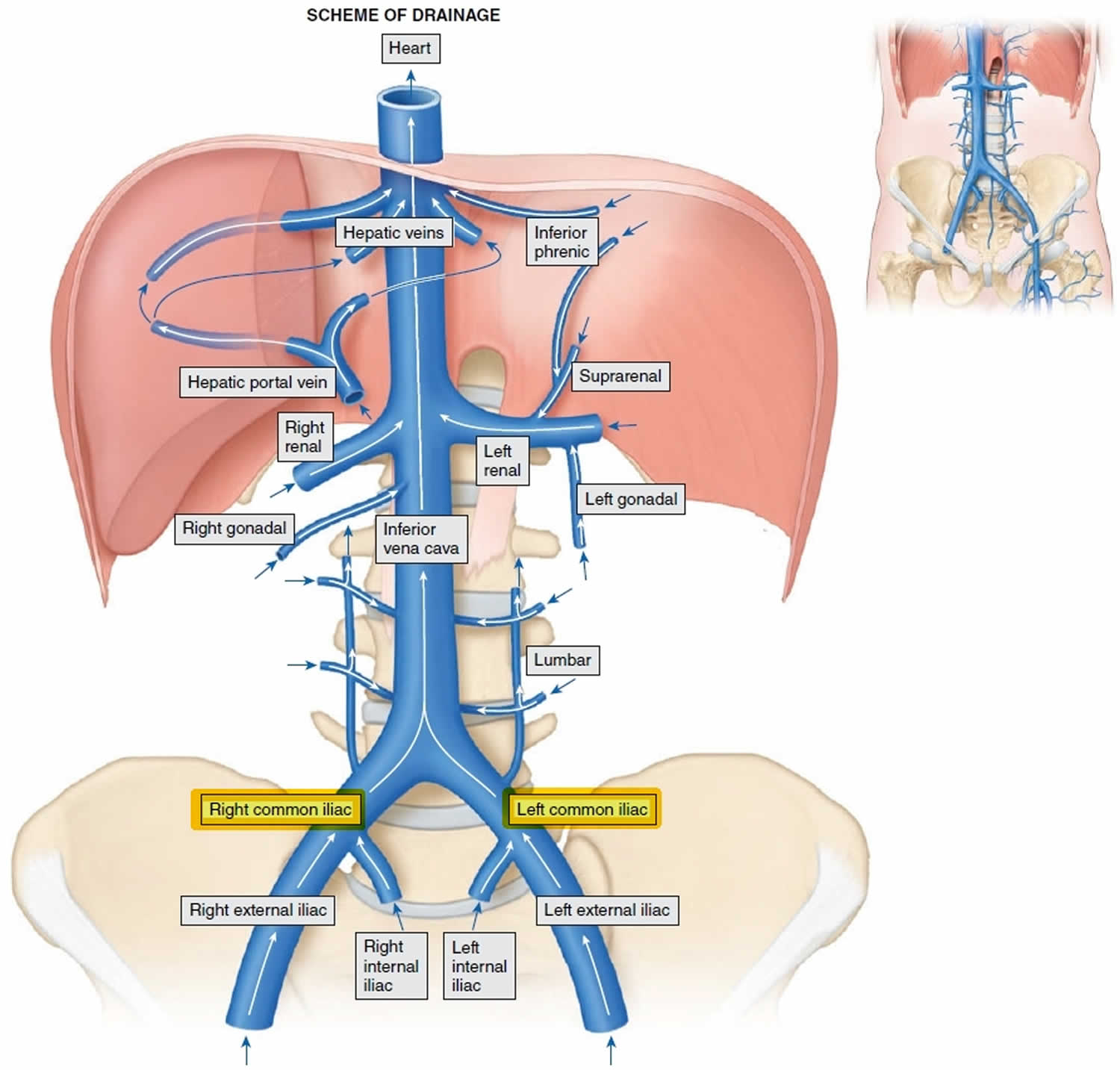
Common iliac vein anatomy and function
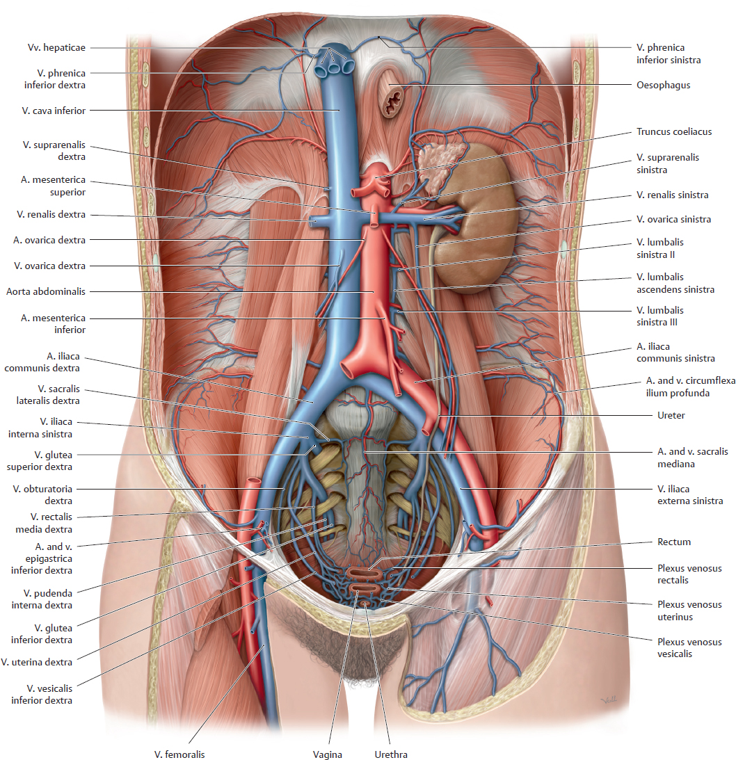
Common iliac vein In human anatomy, the common iliac veins are formed by the external iliac veins and internal iliac veins. The left and right common iliac veins come together in the abdomen at the level of the fifth lumbar vertebra, [1] forming the inferior vena cava. They drain blood from the pelvis and lower limbs.
Iliac arteries Branches and clinical points Kenhub
Arteria iliaca communis Autor: Sophie Eckert • Geprüft von: Claudia Bednarek, Ärztin Zuletzt geprüft: 30. Oktober 2023 Lesezeit: 5 Minuten Die paarige Arteria iliaca communis (gemeinsame Beckenarterie) liegt retroperitoneal in der Bauchhöhle und entsteht aus der Bauchaorta (Aorta abdominalis).. Sie versorgt mit ihren Ästen die untere Extremität, das kleine Becken sowie Teile der Rumpfwand.

Arteria iliaca communis via medici Anatomie lernen, Anatomie, Mensch körper
The common iliac artery (CIA) is a short artery transporting blood from the aorta towards the pelvic region and lower extremity. Sometimes this paired artery is also referred to as its plural form common iliac arteries. The left and right common iliac arteries are the terminal branches of the abdominal aorta.
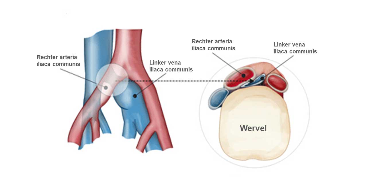
Linkszijdige diepe veneuze trombose door het iliocavaal compressiesyndroom FocusVasculair
Das bilaterale Aneurysma der Arteria iliaca commonis (AIC) ist im Vergleich zum abdominalen Aortenaneurysma seltener, sein Verlauf dagegen ist häufiger symptomatisch. Bei Patienten mit schweren kardiopulmonalen Begleiterkrankungen sind deshalb transluminale und/oder endovaskuläre Vorgehen zur Behandlung des bilateralen Aneurysmas der AIC bevorzugt. Die transluminale bilaterale Okklusion der.
Vena iliaca interna Anatomie, Verlauf & Klinik Kenhub
The common iliac arteries (CIAs) enter the pelvis on the medial aspect of the psoas muscle. The left CIA is shorter than the right. The right CIA passes anterior to the left common iliac vein and then anterior and parallel to the right common iliac vein. The left CIA course is simpler, running parallel and lateral to the left common iliac vein.

Was sind die ersten Äste der Aorta abdominalis für die... Anatomie Abdomen Repetico
The common iliac vein (CIV) (TA: vena iliaca communis), corresponding with the common iliac artery, drains venous blood from the pelvis, lower limbs and their associated structures. Summary location: pelvis, anterior to the sacroiliac joint or.

iliaca interna Leichter lernen, Medizin, Anatomie
Verschlussprozesse an der A. iliaca interna sind häufig symptomfrei, da ein ausgedehntes Kollateralsystem zur Gegenseite sowie zu femoralen, lumbalen und mesenterialen Gefäßen besteht. Klinisch wird die Erkrankung meist durch eine Gefäßclaudicatio und/oder vaskuläre Impotenz symptomatisch.
Left common iliac artery (Arteria iliaca communis sinistra) Kenhub
A. iliaca communis dx. et sin. vznikají rozvětvením břišní aorty Oblast zásobení A. iliaca communis vysílá jen drobné větve do m. psoas major, k mízním uzlinám a k ureteru . Průběh a větvení A. iliaca communis dextra přechází kolmo přes levou v. iliaca communis a hned poté pokračuje před pravostrannou v. iliaca communis.